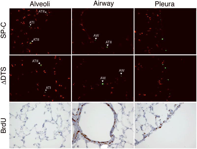
Figure 5. Identification of lung cells expressing YFPnuc and BrdU incorporation.
Plasmids (50 μg) expressing YFPnuc from the CMViep and carrying the SP-C DTS downstream of YFPnuc were delivered to the lungs of Balb/c mice (n=3) by transthoracic electroporation (8 pulses of 10 msec duration at 200 V/cm). The mice were also injected with BrdU (1 mg) 24 and 1 hour prior to gene delivery and 1, 24, and 45 hours after gene delivery. Two days after gene delivery, lungs were harvested and embedded in OCT for frozen sections. Cryosections containing YFPnuc positive cells (green) following BrdU injections, intratracheal delivery and electroporation were counterstained with an ATII-specific antibody, LB180 (red). Separate sections were also reacted with an anti-BrdU antibody and counterstained with hematoxylin. Representative images of specified lung areas are shown.

