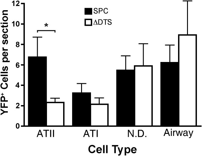
Figure 6. Classification and quantification of lung cells expressing YFPnuc.
Cryosections (15 per animal; spanning the whole lung) containing YFPnuc positive cells following intratracheal delivery and electroporation were counterstained with an ATII-specific antibody. Each cell expressing YFPnuc was defined as an ATII, ATI, airway, or unclassified cell. ATII cells were identified by positive antibody marker staining. ATI cells were identified by location in the lung and a flat nuclear morphology. Airway cells were identified by location in the lung. Cells expressing YFPnuc in the periphery were not scored. (*) indicates statistical differences with p<.05 by a paired one-tail t-test. Data are plotted as mean ± st. dev. from three or more animals.

