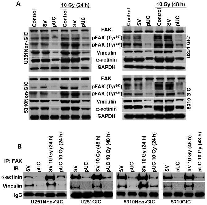
Figure 5.
Depletion of uPAR and cathepsin B inhibits PKC/integrin signaling to FAK and the cytoskeletal molecules. A, Cell lysates from U251 and 5310 non-GICs and GICs were extracted using RIPA buffer after treatment with pUC and radiation alone or in combination, and western blot analysis was performed to determine the expression levels of FAK, pFAK (Tyr397), pFAK (Tyr925), vinculin, and α-actinin using their specific antibodies. GAPDH was used as a loading control. B, The total cell lysates of U251 and 5310 non-GICs and GICs as described above were immunoprecipitated with FAK antibody (2 μg). The protein precipitates were washed with lysis buffer and incubated with 1X loading dye at 90°C for 10 min. SDS-PAGE was conducted and western blotting was performed with vinculin and α-actinin antibodies as described in Materials and methods.

