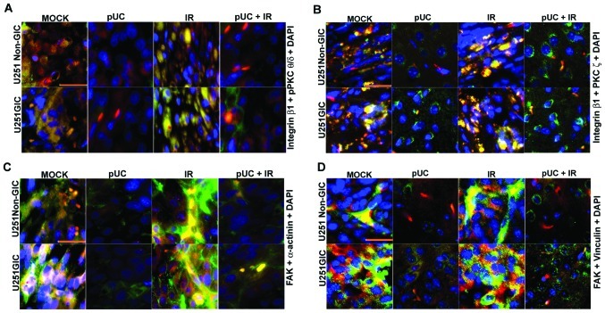
Figure 7.
Effect of pUC and radiation treatment on pre-established intracranial tumors. U251 non-GICs and GICs were implanted intracranially into nude mice and treated with 450 μg of pUC seven days after implantation. Radiation treatment was given in two doses (5 Gy at days 8 and 10). When chronic symptoms were observed, the mice were sacrificed, and their brains were collected and embedded in paraffin. Paraffin-embedded sections were deparaffinized, antigen retrieved, and co-localization studies were carried out. The brain sections were incubated overnight with primary antibodies in 10% goat serum at 4°C in a humidified chamber, counterstained with Alexa Fluor-conjugated secondary antibodies, and incubated with DAPI for nuclear staining before mounting. A, Co-localization of integrin β1 (green), pPKC θ/δ (red) and DAPI (blue) in the paraffin sections of the nude mice established with U251 non-GICs and GICs and treated with shRNA and radiation alone or in combination. B, Interaction between integrin β1 (green) and PKC ζ (red) in the various in vivo combination treatments. DAPI (blue) was used for nuclear staining. C, In vivo brain sections of immunocompromised mice implanted with U251 non-GICs and GICs were labeled with FAK (green) and α-actinin (red) and processed for immunofluorescence. The sections were incubated with DAPI for a brief period of time for nuclear staining. D, Mock, pUC-treated, irradiated, and pUC + irradiated brain sections of the nude mice pre-established with U251 non-GICs and GICs were immunoprocessed and labeled with FAK (green), vinculin (red) and DAPI (blue) in order to observe the co-localization of FAK and vinculin in in vivo samples.

