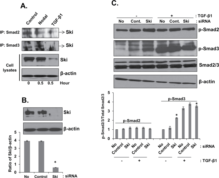
Fig. 5.
Roles of Ski in Nodal and TGF-β signaling in prostate cancer cell lines. (A) PC3 cells were treated with Nodal (200ng/ml) and TGF-β1 (5ng/ml) for 0.5h. Proteins were immunoprecipitated from cell lysates using anti-Smad2/3 antibodies followed by western blot analysis with anti-Ski antibody. Total cell lysates were also collected to determine the expression of Ski and β-actin (internal control). (B) Knockdown of endogenous Ski protein in PC3 cells by specific Ski siRNA. Equal amount of cell lysates were analyzed by western blot using anti-Ski antibody (upper panel). The levels of β-actin in cell lysates were used as internal controls. Quantitative analysis of PC3 cells with or without Ski siRNA or control siRNA after normalization to the signal obtained with β-actin (lower panel). * Significantly different (P < 0.05) from cell transfected with control siRNA. (C) Western blot analysis of phosphorylated Smad2 and Smad3, total Smad2/3 and β-actin in PC3 cells transfected with control or Ski siRNA. The cells were incubated in the presence or absence of TGF-β1 (5ng/ml) for 10min. Western blots using anti-Smad2/3 and β-actin antibodies as internal controls. Quantitative analysis of p-Smad2 and p-Smad3 in PC3 cells with or without treatment with TGF-β1 (5ng/ml) (lower panel) were relative to untreated control (designated as one) after normalization to the signal obtained with Smad2/3. Each bar represents mean ± SEM (n = 3). *Significantly different (P < 0.05) compared with untreated controls.

