Figure A1.
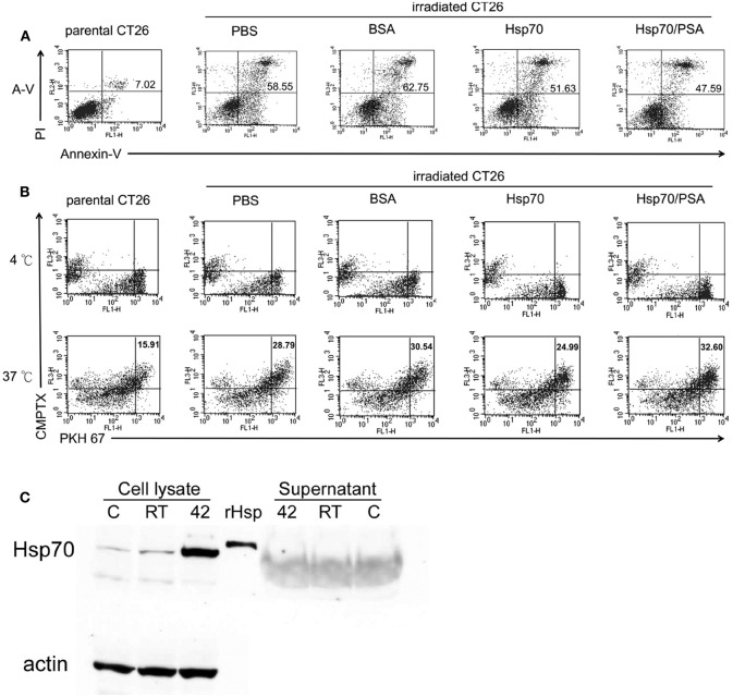
Exogenous rHsp70 did not increase radiation–induced CT26/PSA apoptosis nor increase BM-DC phagocytosis. (A) The percentage of apoptotic CT26/PSA cells was analyzed by annexin-V and PI (propidium iodide) staining (shown in the upper right corner) in, left to right, parental CT26/PSA cells in PBS (parental CT26) or irradiated CT26/PSA cells (irradiated CT26) in PBS (PBS) or 10 μg/ml of BSA (BSA), rHsp70 (HSP70), or rHsp70C′-PSA (Hsp70/PSA) for 24 h incubation. The number of percentage was indicated AnnexinV-positive cells. (B) Cell Tracker-labeled CT26/PSA (5 × 10⁵), pretreated as in Figure 1A, and PKH-67-labeled BM-DCs (5 × 10⁵) were cocultured in 6-well plates for 24 h at 4°C (top row, no phagocytosis control) or 37°C (bottom row) and the percentages of double-positive cells measured. The panels are arranged in the same order as in Figure 1A. (C) Untreated (lane C) or irradiated CT26/PSA cells (lane RT) or control cells heated at 42°C for 30 min (positive control; lane 42) were harvested for Western blot analysis for Hsp70 protein in the cell lysate and culture supernatant. Lane rHsp is rHsp70. These data are representative of those obtained in three experiments.

