
Figure A2.
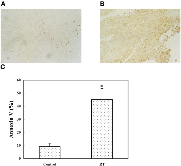
In vivo tumor apoptosis assay. CT26/PSA cells (1 × 10⁵) were injected s.c. on day 0. On day 14, the tumor size was around 4 mm in diameter and the mice were left untreated (A) or received 8 Gy of RT (B), then, on day 16, sections from the tumor site were analyzed using the TUNEL assay. Apoptotic cells are stained brown (magnification, 100 ×). (C) The tumor mass was digested with collagenase and analyzed by annexin V staining; the apoptotic cells are expressed as the percentage of annexin V-positive cells. The data represent the mean ± SD for three mice. *Indicates p < 0.05.