Fig. 5.

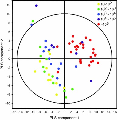
Scores plot of the PLS model of urine 1H NMR spectra at baseline versus the number of bacteria (CFU/ml) found in urine (R2Y = 0.78, Q2 = 0.44). Colored by the number of bacteria (Color figure online)

Scores plot of the PLS model of urine 1H NMR spectra at baseline versus the number of bacteria (CFU/ml) found in urine (R2Y = 0.78, Q2 = 0.44). Colored by the number of bacteria (Color figure online)