Figure 11.
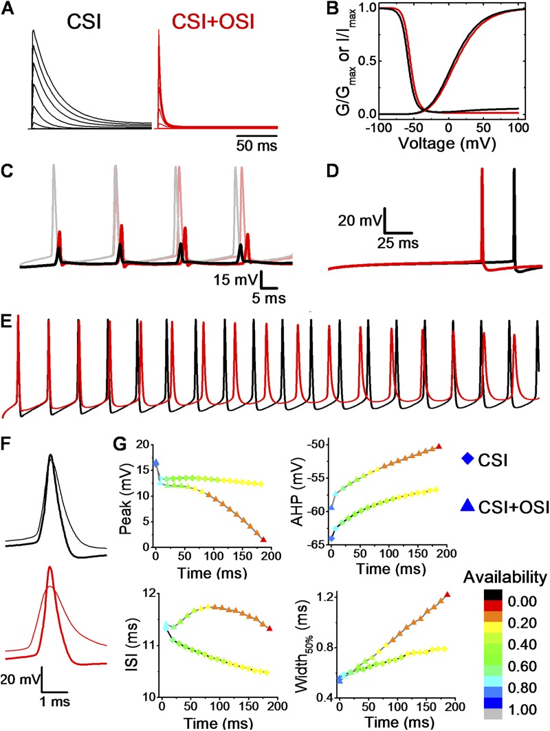
The pathways of Kv4 channel inactivation influence spiking properties in a computational model of the CA1 pyramidal neuron. (A) NEURON simulation of current families evoked by depolarizations from −60 to +80 mV (20-mV steps). Holding potential was −100 mV. (B) Gp-V and steady-state inactivation curves of CSI (black) and CSI + OSI (red). (C) Somatic (dim traces) and the corresponding dendritic (solid; 400 µm from the soma) APs from a 90-ms stimulation of 150 pA at the soma. (D) Latency to the first somatic spike with a 40-pA stimulation for 200 ms (rheobase for CSI model at 200 ms). (E) Simulated somatic AP trains assuming specific Markov schemes: CSI (black) and CSI + OSI (red). AP firing was elicited by injecting 250 pA over a period of 200 ms. (F) Overlay of the first (thick) and the last (thin) AP in the trains shown in A. (G) Summary of somatic AP properties. Each point corresponds to an AP, and the shape of the symbol denotes the pathway of inactivation (diamond, CSI; triangle, CSI + OSI). The color gradient represents the fraction of available Kv4.2 channels at the AHP before the spike (i.e., maximal availability preceding the spike).

