Figure 6.
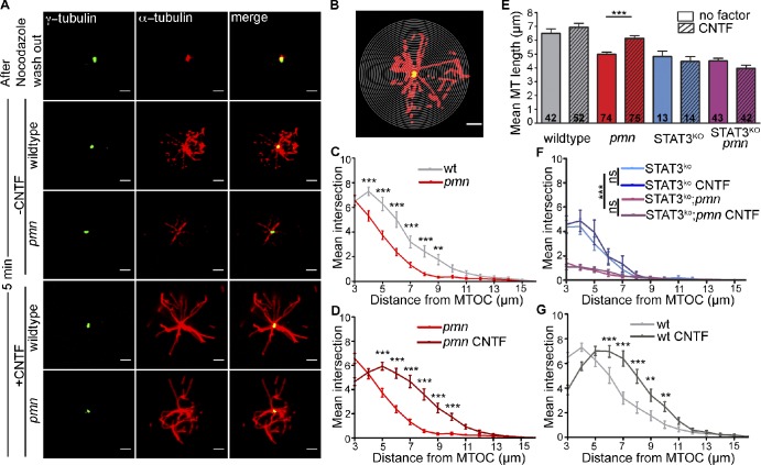
CNTF enhances MT regrowth in cultured motoneurons. (A) MTs were depolymerized with nocodazole, and MT regrowth was analyzed at 5 min after CNTF application in cultured wild-type and pmn mutant motoneurons. The centrosome is labeled with γ-tubulin (Cy2), and MTs were labeled with α-tubulin (Cy3). Bars, 2 µm. (B) Representative image from Sholl analysis performed on primary motoneurons with 0.25-µm step concentric circles. Bar, 2 µm. (C, D, F, and G) Graphs obtained from Sholl analysis depicting number of intersections on y axis and distance from MTOC on the x axis. (C) Comparison of MT regrowth between pmn mutant and wild-type motoneurons. (D) CNTF enhances MT regrowth in pmn mutant motoneurons. (E) Graphical representation of mean length of polymerized MTs formed in wild-type and pmn mutant motoneurons with and without CNTF and in STAT3fl/KO;NFL-Cretg motoneurons. Numbers in bars represent the number of analyzed motoneurons. Error bars shown represent means ± SEM from four independent experiments. Statistical analysis: ***, P < 0.001; ANOVA with Bonferroni posthoc test. (F) CNTF-mediated MT regrowth was abolished in STAT3fl/KO;NFL-Cretg and STAT3fl/KO;NFL-Cretg;pmn mutant motoneurons. (G) CNTF enhances MT regrowth in wild-type motoneurons. Statistical analysis: **, P < 0.01; ***, P < 0.001; two-way ANOVA with Bonferroni posthoc test. wt, wild type.

