Figure 4.
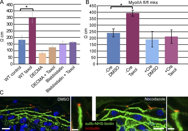
Tight junction barrier activity is increased by cortical microtubules. (A) Transepithelial resistance (TER) measurements were taken of wild-type cells, cells treated with taxol, cells treated with E-cadherin inhibitory DECMA antibodies (with and without taxol), and in blebbistatin-treated cells (with and without taxol). P = 0.0037 for control vs. taxol treatment; n = 9. (B) TER levels in myosin IIA WT and null cells (with and without taxol). P = 0.007. (C and D) In vivo epidermal barrier assay. DMSO or nocodazole was injected subcutaneously with a biotin tracer. After 30 min the skin was removed, embedded, and sectioned for analysis. Streptavidin-FITC (green) allowed visualization of the diffusion of the biotin, and occludin (red) puncta mark the tight junctions in the granular layer of the epidermis. Bar, 10 µm (5 µm for insets).

