Figure 6.
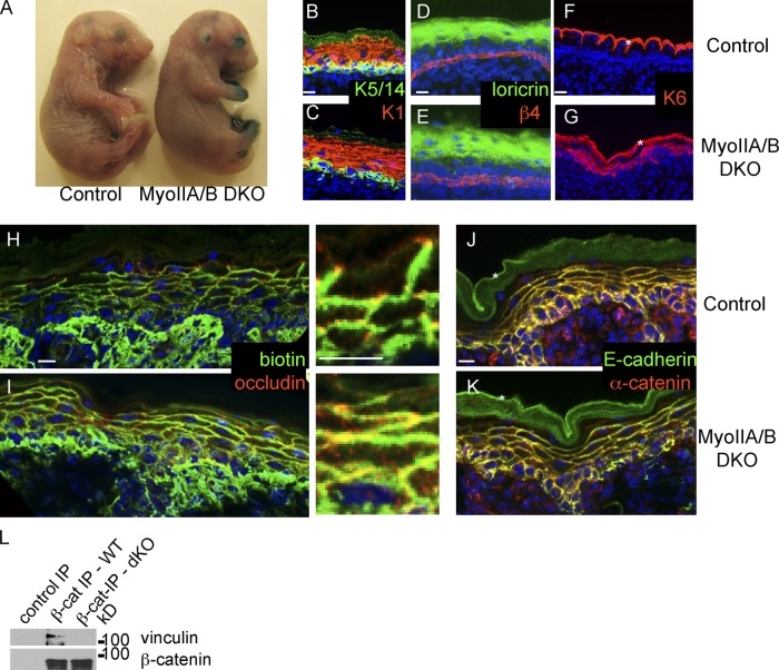
Loss of myosin IIA and B results in tight junction defects in the epidermis. (A) Barrier assay in E18.5 control littermate (left) and myosin IIA/B double cKO epidermis. Note the barrier defects on digits, eyes, and ears, and normal barrier function over the rest of the body. (B and C) Normal expression of differentiation markers: K5/14 (green) and K1 (red) in control (B) and myosin II dKO epidermis (C). (D and E) Normal expression of granular layer marker loricrin (green) in control (D) and myosin II dKO epidermis (E). β4-integrin (red) marks the basement membrane in these images. (F and G) Abnormal expression of the stress marker keratin 6 (K6, red) in myosin II dKO epidermis (G). Asterisks mark nonspecific cornified envelope staining in F and G. (H and I) Biotin diffusion assay in control (H) and myosin II dKO (I) epidermis. Biotin is detected with streptavidin (green) and tight junctions are marked with occludin (red). (J and K) Adherens junction components E-cadherin (green) and α-catenin (red) in control (J) and myosin II dKO epidermis (K). (L) Extracts from Myosin IIA/B dKO epidermis or littermate controls were immunoprecipitated with anti–β-catenin antibodies. Bound proteins were analyzed by Western blotting with vinculin and β-catenin. Bars, 10 µm.

