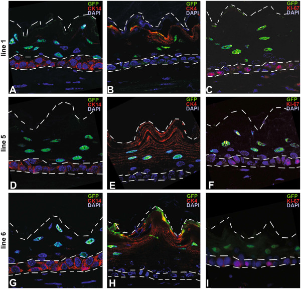
Figure 2.
H2B-GFP expressing cells are located exclusively within the differentiated non-basal cell layers. Immunofluorescence double staining for GFP (green, nuclear) and A,D,G) CK14 (red, basal cell layer), B,E,H) CK4 and C,F,I) CK13 (both red, non-basal cell layers) in all three founder lines (A-C line 1; D-F line 5, G-I line 6, all 40x magnification). The dashed lines indicate the outline of the esophageal epithelium and the border to the basal cell layer.

