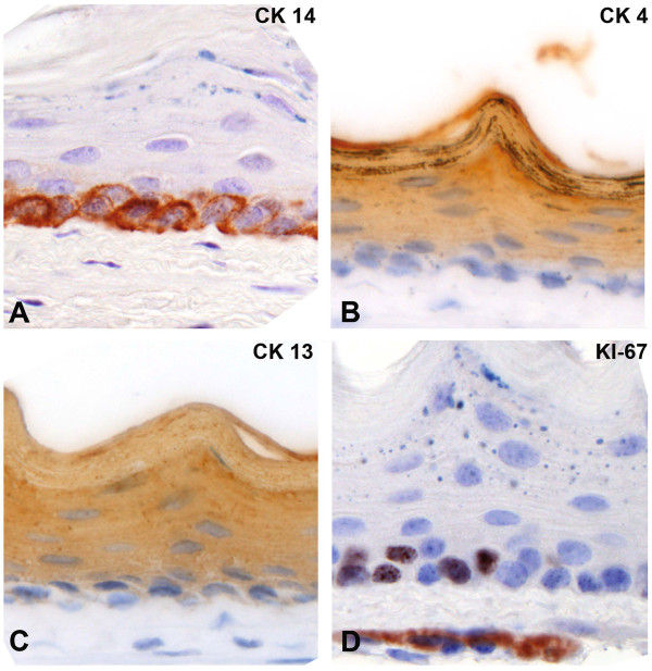
Figure 3.
IHC analysis of the squamous epithelium of the mouse esophagus. Immunohistochemistry for cytokeratin 14 marks the basal layer (A), while cytokeratin 4 and 13 are expressed in the differentiated cell layers above the basal cells (B, C). The proliferation marker Ki-67 is expressed in distinct basal cells (D). All images were taken at 40x magnification.

