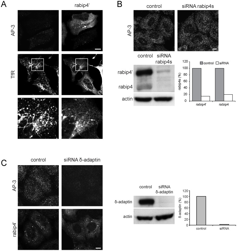
Figure 7. Rabip4’ localization does not require AP-3.
VSVG-rabip4’-expressing HeLa cells were treated with 5 µg/ml BFA for 15 min at 37°C and stained with a rabbit antibody against rabip4’ and mouse anti-δ-adaptin or mouse anti-TfR, followed by Alexa568-anti-rabbit and Alexa488-anti-mouse IgG. Rabip4’ overexpression did not affect AP-3 sensitivity to BFA. Lower row represents insets of boxed areas. Arrows point to BFA-induced tubulation of rabip4’ and TfR (A). Rabip4s-directed siRNA oligos were transfected in HeLa cells for 3 days. AP-3 distribution was similar in both siRNA-transfected and control cells. Scale bar is 10 µm. Silencing was monitored by Western blotting and gave routinely 80–85% reduction of both rabip4’ and rabip4 isoforms (B). AP-3 siRNA oligos were transfected in HeLa cells for 3 days. Two days after siRNA treatment, cells were transfected with VSVG-rabip4’ for another day and labeled for δ-adaptin and rabip4’. Rabip4’ distribution did not depend on AP-3. Scale bar is 10 µm. Western blots were probed with antibodies against δ-adaptin. The level of δ-adaptin in siRNA-transfected cells was quantified and expressed as % of control (C).

