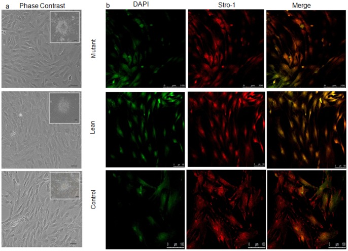
Figure 1. Characterization of the primary cultures of BM-MSCs.
(A) Phase contrast photomicrographs showing primary cultures of BM-MSCs (day 7) from Mutant, Lean and Control. Spindle-like cell morphology with colony-formation units (CFU) were seen (insight), which have been captured using ACT2U software attached to Nikon Microscope at magnifications represented using scale bar. (B) MSCs characterized for mesenchymal-specific cytosolic protein STRO-1 (red) and cell nuclei were stained with 4′,6-diamidino-2-phenylindole (DAPI) (green - pseudo color). Images were captured in Confocal Microscope using Leica Advanced Fluorescence software (Leica SP5 series, Germany) at magnifications represented using scale bars.

