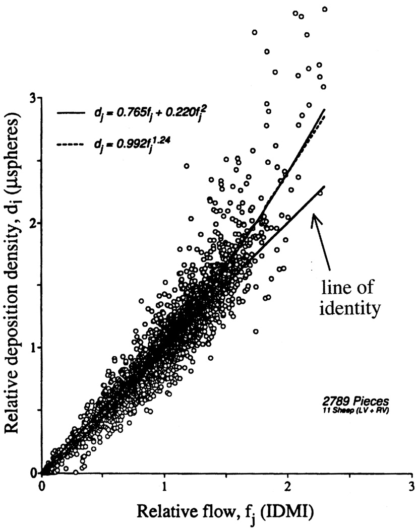
Fig. 1.
Microsphere deposition densities versus 2-iododesmethylimipramine (IDMI) deposition densities in ventricular myocardium of 11 open-chest sheep. The best fitting linear regression line was di = 1.268fi − 0.255, r = 0.925; the best fitting quadratic through the origin (- - -, r = 0.945) and best power fit (———, r = 0.948) are presented. The line of identity is given as a reference. Piece sizes averaged about 220 mg. The relatively higher deposition of microspheres relative to that of the molecular marker IDMI in high flow regions is more than can be accounted for by incompleteness of IDMI extraction and demonstrates a small bias for microspheres to flow in arterioles with higher flow. (Figure reproduced from Ref. [14] with permission from the American Heart Association.)

