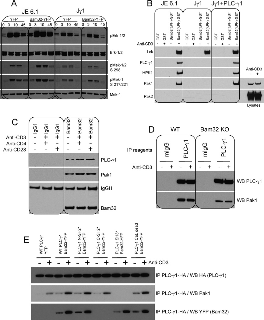
Figure 2. Bam32 interacts with Pak1 via PLC-γ1 and Bam32-PLC-γ1-Pak1 cooperative complexes increase Mek-1 and Erk phosphorylation.
(A) JE6.1 and Jγ1 cells transfected with YFP or Bam32-YFP cDNAs were sorted for equal YFP fluorescence intensity. Cells ± αCD3 were analyzed by WB. A representative WB is shown (n=4).
(B) JE6.1, Jγ1 and Jγ1 cells expressing PLC-γ1 ± αCD3 were lysed. Extracts (1 mg total protein) were incubated with GST or Bam32(ΔPH)-GST proteins. Bound proteins were analyzed by WB. For Pak1 and Pak2 analysis WCL from resting and OKT3-stimulated Jγ1 cells were loaded to confirm Pak2 expression in Jγ1. A representative WB is shown (n=2).
(C) Human CD4+ T cells were stimulated with αCD3 (5 µg/ml) + αCD4 (10 µg/ml) or αCD28 (10 µg/ml). Lysates were subjected to a Bam32 or control IP. IPs were analyzed by WB. A representative WB is shown (n=2).
(D) CD4+ T cells from WT or Bam32−/− mice were stimulated with αCD3ε (1 µg/ml, 2 min.). Lysates were subjected to a PLC-γ1 or control IP. IPs were analyzed by WB. A representative WB is shown (n=4).
(E) JE6.1 cells were transfected with YFP or WT Bam32-YFP + WT PLC-γ1-HA or mutants of PLC-γ1-HA (10 µg each). Lysates from cells ± αCD3 stimulation were subjected to an HA IP to pull down PLC-γ1. A representative WB is shown (n=3).

