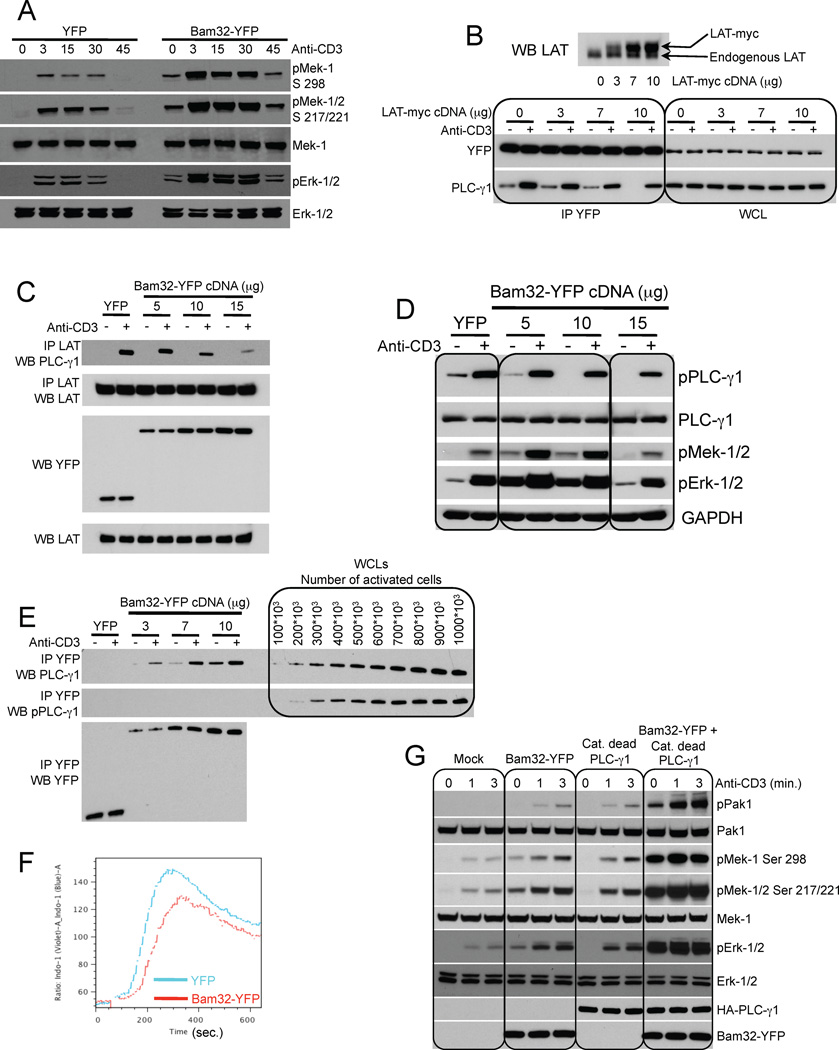
Figure 4. The Bam32 pathway regulates Erk activation independently of the canonical LAT pathway.
(A) JCam 2.5 cells stably transfected with either YFP or Bam32-YFP cDNAs were sorted for the same YFP intensity. Cells were stimulated or not with αCD3. WCL were analyzed by WB. A representative WB is shown (n=2).
(B) LAT-myc cDNA was overexpressed in JE6.1 cells stably expressing Bam32-YFP. Lysates from cells ± αCD3 were subjected to a YFP IP. WCL were run to monitor the input. Upper panel is a LAT WB showing the increase of LAT-myc in addition to endogenous LAT. A representative WB is shown (n=2).
(C, D) JE6.1 cells were transfected with YFP or increasing amounts of WT Bam32-YFP. Cells ± αCD3 were lysed. Lysates were split in half. Half of the lysates were subjected to a LAT IP (panel C) and the other half was used to make WCL (panel D). Samples were analyzed by WB. Representative WBs are shown (n=2).
(E) JE6.1 cells were transfected with YFP (7 µg) or increasing amounts of WT Bam32-YFP cDNAs. Cells ± strong αCD3 (10 µg/ml) were lysed. Lysates were subjected to a Bam32-YFP IP. The amount of PLC-γ1 bound to Bam32-YFP and its phosphorylation state were studied by WB. In parallel, JE6.1 cells were stimulated under the same conditions and lysed. Increasing amounts of this WCL were loaded on the same gel. A representative WB is shown (n=2).
(F) JE6.1 cells were transfected with YFP or WT Bam32-YFP cDNAs (10 µg). Cells loaded with Indo-1 AM were stimulated with αCD3 and cytosolic Ca2+ concentration was assayed by flow cytometry. Data are representative of 4 independent experiments.
(G) JE6.1 cells were transfected with WT Bam32-YFP, catalytically dead PLC-γ1 or both (7.5 µg). Cells were stimulated or not with αCD3 (500 ng/ml, 3 min.). WCL were analyzed by WB. A representative WB is shown (n=2).

