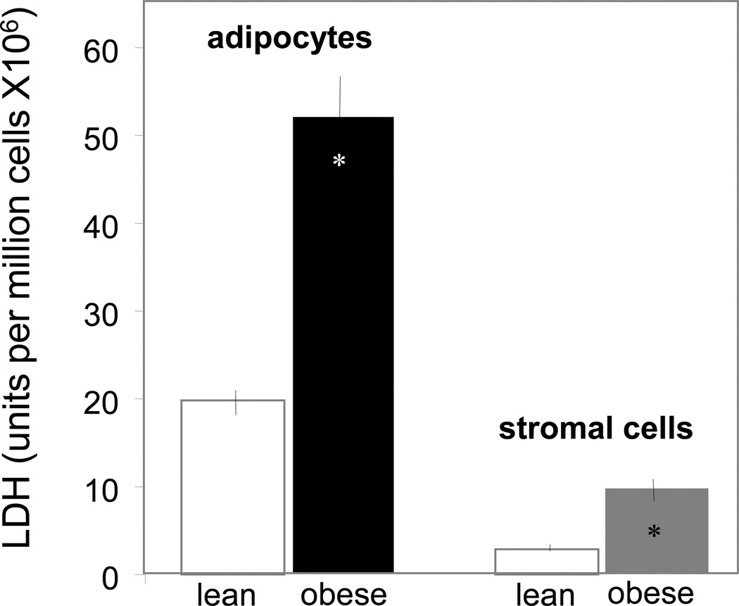
Figure 3. Biochemical analysis of necrosis in adipose cells of obese pregnant women.
Necrosis was estimated by the amount of lactate dehydrogenase released during a 24 h period in the incubation medium of adipocytes and cells of the stromal vascular fraction. Cells isolations were independently performed from adipose tissue of 10 lean and 15 obese pregnant women. Results expressed as mean ± SD.

