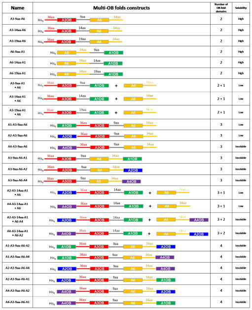
Fig. 1. Linked OB-folds and expression results.
A schematic representation of the multi-OB-fold domain constructs is shown in the second column with the A1 in green, A2 in blue, A3 in red, A4 in purple, A6 in yellow and flexible GS repeat linkers in grey. OB-fold domains and connecting linkers are shown as boxes and solid lines, respectively. Lengths of linker peptides are indicated on the top of solid lines. Recombinant proteins of both fused and separate multi-OB-fold domains were characterized by size exclusion chromatography, as summarized in Table 1. The symbol + indicates that two proteins were co-expressed from different plasmids (see Methods).

