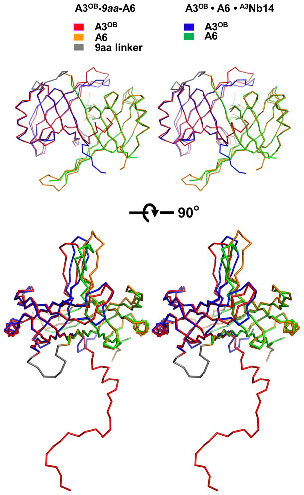
Fig. 3. Comparison of A3OB and A6OB dimers from the A3OB-9aa-A6 and A3OB•A6•(A3Nb14)2 structures.
Backbone superpositions of the A3OB and A6OB domains from the current A3OB-9aa-A6 and A3OB•A6•(A3Nb14)2 structures (PDB-ID: 3STB; (Park et al., 2012a)) are shown. The A3OB domain of A3OB-9aa-A6 is in red; the A6OB domain of A3OB-9aa-A6 in yellow; the A3OB domain of A3OB•A6•(A3Nb14)2 in blue, and the A6OB domain of A3OB•A6•(A3Nb14)2 in green. The core structures of the anti-parallel β-barrel in both fused and separate dimers are almost identical with a root-mean-square deviation of 0.93 Å.

