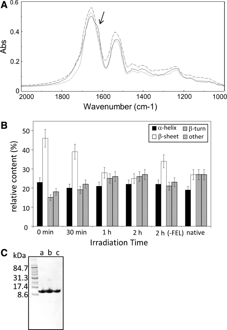
Fig. 2.
Effect of FEL irradiation on the structural change of lysozyme fibrils. A FTIR spectra of lysozyme samples before and after the FEL irradiations at 1,620 cm−1 for 2 h. The measurements were operated using KBr-plate and 16 scans. Solid line the spectrum of the enzyme before the irradiation; dotted line that of the enzyme after the irradiation; dashed line that of native enzyme. An arrow indicates the wave number targeted by the FEL irradiation. B Secondary-structure analyses. The percentages were calculated based on the de-convoluted spectra of amide I bands. “Others” indicate the disordered region. The experiments including FEL irradiation and the following measurement of FTIR spectra were performed five times, and the statistical errors of secondary-structural changes, which were mainly caused by the FEL irradiation conditions, were evaluated to be ±10 % (one standard deviation). C SDS-PAGE analyses. The lysozyme samples were loaded on the gel (15 %) without heat denaturing. a native lysozyme, b fibrils, c fibrils following the FEL irradiation

