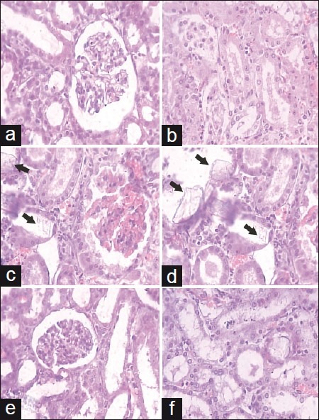
Figure 2.

Light microscopic histology and CaOX deposits in the kidney section. Kidney section of (a, b) vehicle control, (c, d) urolithic and (e, f) cystone treated (750 mg/kg). (a: glomeruli region and b: tubular region) (magnification 450× for all images)
