
Figure 3.
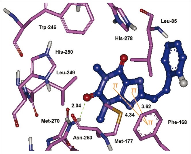
The lowest energy configuration of docking result of caffeinyl analog (Compound 10) with binding pocket of human AA2AR. The residues of binding pocket are shown as stick in pink color while compound 10 is presented as ball and stick style in blue color. Dashed lines in green indicate H-bonds while π–π stacking interaction are shown as orange lines. Sulfur is presented in dark yellow and oxygens in red