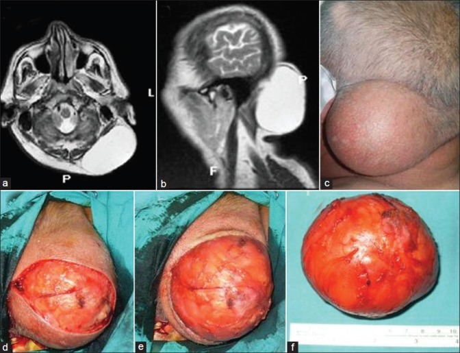
Figure 2.

(a) Axial and (b) sagittal T2-weighted MR imaging shows the giant lipoma; (c) the lipoma in the occipital region; (d) the capsule of the lipoma after skin incision; (e) the resected lipoma peeled off from the surrounding tissue and removed together with the capsule; (f) picture of the completely resected lipoma
