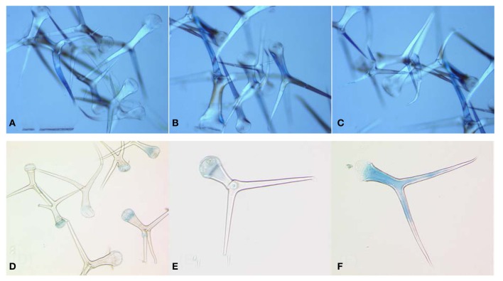
Figure A1.
Microscopic image of isolated trichomes. (A–C) show three different biological replicates of independent trichome isolations. (D–F) Analysis of trichom intactness after trypan blue staining; (D) Pool of isolated trichomes after trypan blue staining; (E) Intact trichome. Trypan blue is only visible on the base of trichome where it was attached to the epidermal cells; (F) shows damaged trichome. Cytoplasm of cell is stained in blue because of destruction of inner membranes and dye penetration into the cell. About 2–5% of trichomes showed visible staining of the cell cytoplasm.

