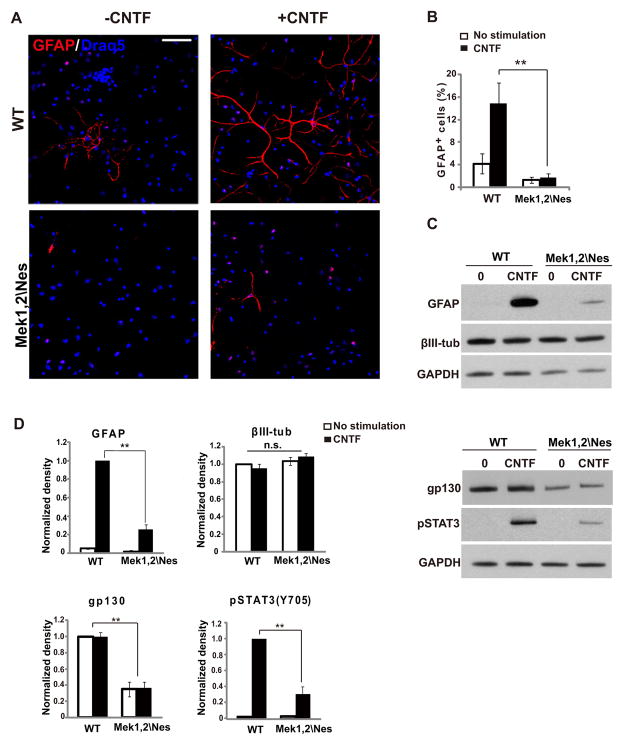
Figure 4. Gliogenic signaling is impaired in Mek1/2 deleted progenitor cultures.
(A) Representative images of cortical progenitor cultures derived from the E17.5 Mek1,2\Nes mutant and WT dorsal cortices. Cultures were stimulated with CNTF for 5 days and immunolabeled for GFAP (red) and Draq5 (blue). CNTF did not induce substantial levels of astrocyte differentiation in mutant cultures. Scale bar= 50um. (B) Quantification of the percentage of GFAP+ cells after 5 days of culture (mean ± s.e.m; n=4(>500 cells); ** = p-value < 0.01, paired t-test). (C) Upper panel: Western blotting revealed strong reductions in GFAP expression, while expression of the neural marker, βIII-tubulin, was not affected in mutant cultures when compared to WT. Progenitors were cultured for 3 days with CNTF as indicated. Lower panel: Further analysis of CNTF stimulated progenitor cultures by Western blotting demonstrated that gp130 expression and phosphorylated STAT3 levels in E17.5 Mek1,2\Nes mutant culture were profoundly reduced when compared to WT. Progenitors were cultured for 3 days prior to treatment with CNTF for 15 minutes as indicated. (D) Quantification for levels of GFAP, βIII-tubulin, gp130 and pSTAT3 in (C). (Mean ± s.e.m; n=3; ** = p-value < 0.01; n.s.: not statistically significant; paired t-test).

