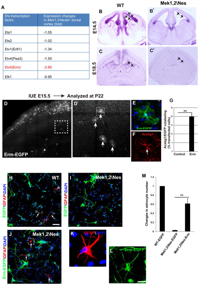
Figure 5. Erm promotes glial progenitor specification and rescues gliogenesis of Mek deleted progenitors in vitro.
(A) Microarray analysis of E18.5 WT and Mek1,2\Nes mutant cortices showed that Etv5 (Erm) mRNA expression was strongly reduced in mutants, while other Ets family members were changed to a lesser extent. (B–B′) In situ hybridization revealed that Erm is predominantly expressed in VZ of E14.5 WT cortex (arrows); however, Erm expression in Mek1,2\Nes VZ was profoundly reduced. (C–C′) Erm is expressed broadly in E18.5 WT dorsal cortex VZ (arrows), hippocampus, and deep cortical layers. In mutant brains, Erm expression was dramatically downregulated in the VZ but not in deep cortical layers. (D–D′) Overexpression of Erm-EGFP in E15.5 radial progenitors promoted a dramatic increase in the number of mature astrocytes in postnatal day 22 dorsal cortices. D′ is the delineated areas in D. Arrows in D′ indicate EGFP labeled astrocytes. (E–F) High magnification images of the astrocyte from the delineated area in D′ that expresses Erm-EGFP and Acsbg1. (G) The proportion of EGFP+ astrocytes as quantified in EGFP or Erm-EGFP transfected cortices. (Mean ± s.e.m; ** = p-value < 0.01; paired t-test, n=4). (H–J) Representative images showing electroporation of radial progenitors ex vivo at E14.5 followed by dissociation and CNTF stimulation for 5 days. Some WT progenitors differentiated into astrocytes (arrows in H). However, EGFP transfected Mek1,2\Nes mutant progenitors did not become astrocytes (I). Electroporation of Erm-EGFP into mutant progenitors rescued astrocyte differentiation (arrows in J). Scale bar=100μm. (K–L) High magnification images from delineated area in (J) clearly show that Erm-EGFP transfected cells expressed GFAP. Scale bar= 20μm. (M) Quantification shows that in Erm transfected mutant cultures, astrocyte number reached 60% that of the EGFP transfected WT cultures. (Mean ± s.e.m; N=4. **=p value<0.01, paired t test). See also Figure S4.

