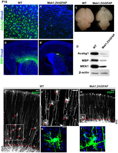
Figure 7. Deletion of Mek1/2 results in persistent disruption of gliogenesis.
(A–B′) In the P10 mutant brain, the number of Acsbg1+ astrocytes and MBP+ oligodendrocytes in dorsal cortices were dramatically reduced. cc: corpus callosum. (C) Mek1,2\hGFAP brains are smaller and appear transparent at P10. (D) Western blots confirmed the dramatic reductions of Acsbg1 and MBP expression in Mek1,2\hGFAP dorsal cortices. (E–G) EGFP was electroporated at P1 to follow maturation of radial progenitors in control and Mek deleted animals. EGFP-expressing mature appearing astrocytes were readily apparent in P8 WT dorsal cortices (arrows in E and F), F is the delineated area in E, G is the delineated area in F. Scale bars=100 μm in E and F, 10 μm in G. (H–I) EGFP transfected Mek1,2\hGFAP cortices. Note that no mature-appearing astrocytes are observed. I is the delineated area in H. EGFP expressing cells remained close to SVZ and did not exhibit typical astrocytic morphology (I). N=4. See also Figure S6.

