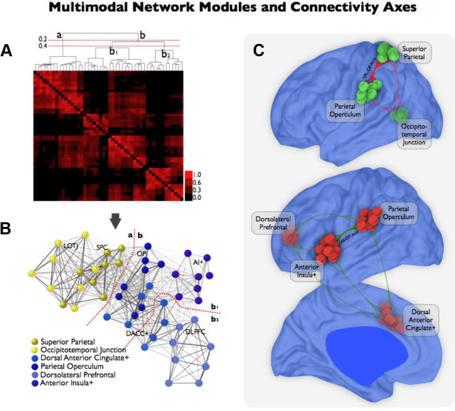
Figure 11.
Modules and connectivity axes of the multimodal integration network. A, B, The average-linkage hierarchical clustering analysis of the multimodal integration network (A) confirmed two main set of regions: (1) module a, formed by superior parietal cortex (dark yellow) and lateral occipitotemporal junction (light yellow), and (2) module b, formed by the parietal operculum (dark blue), anterior insula+ (violet), dorsal anterior cingulate+ (blue), and dorsolateral prefrontal cortex (light blue; B). The dendrogram shows, at a cutoff criterion of r > 0.2, the main partition in a and b modules but also a meaningful subdivision within module b at cutoff criterion r > 0.4. Submodule b1 is formed by parietal operculum (dark blue), anterior insula+ (violet), and part of dorsal anterior cingulate+ (blue), and submodule b2 is formed by dorsolateral prefrontal cortex (light blue) and part of dorsal anterior cingulate+ (blue). C, These results pointed out the existence of major cores and functional connectivity axes within the multimodal integration network. For instance, the superior parietal cortex–parietal operculum connectivity axis was important for the communication between modules a and b. The parietal operculum, anterior insula+, and part of the dorsal anterior cingulate nodes formed a central core in the overall organization of the multimodal network.

