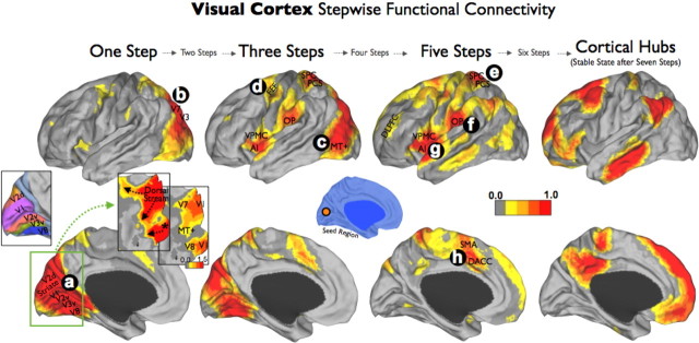
Figure 3.
Visual cortex stepwise functional connectivity. Visual cortex SFC analysis revealed that visual cortex's direct connectivity follows three different pathways, two dorsal (a–c) and one ventromedial (a; inset, star). In addition to the conventional cortical surface and to avoid a ceiling visualization effect in the degree of connectivity of early visual regions, two flat projections centered on the occipital lobe (green square) are presented using the same (top inset) and a relaxed color-scale threshold (bottom inset). In subsequent steps, the visual cortex connectivity reached the frontal eye field (d), the multimodal network (e–h), and finally, the cortical hubs of the human brain. The visuotopic map was provided by Caret software (Van Essen and Dierker, 2007). Visuotopic areas: V1, V2, V3, V7, V8, and MT+; PCS, posterior central sulcus; SMA, supplementary motor area.

