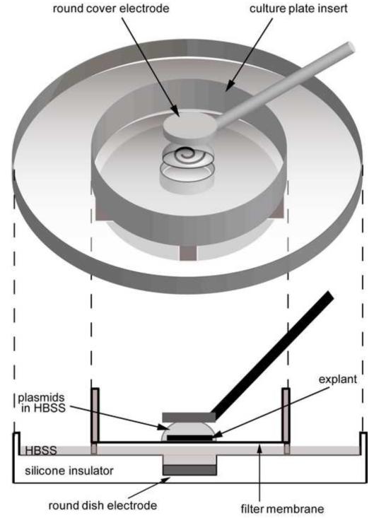
Fig. 3. Electroporation paradigm.
A postnatal day 1.5 (P1.5) cochlear sensory epithelial explant is placed on a filter membrane and covered with 5 μl of HBSS containing a desired plasmid(s). The gap between the filter membrane and the dish electrode is filled with 500 μl HBSS. Five rectangular electrical pulses (12 V, 30 ms duration, 970 ms interval) are applied through the 2 mm diameter round cover (anode) and dish (cathode) electrodes.

