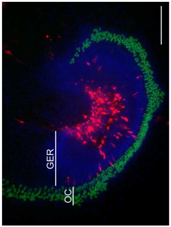
Fig. 4. Typical location of transfected cells in the P1.5 sensory epithelium of the Pou4f3-GFP transgenic mouse.
The explant shown was electroporated with 1 πg/πl dsRed-expression vector, and fixed 2 days after transfection. The majority of the transfected cells are located in the greater epithelial ridge (GER) which was medial to the organ of Corti (OC). As in this example, HCs (expressing GFP) were never transfected. Blue fluorescence shows nuclei labeled with DAPI. The scale bar = 200 μm.

