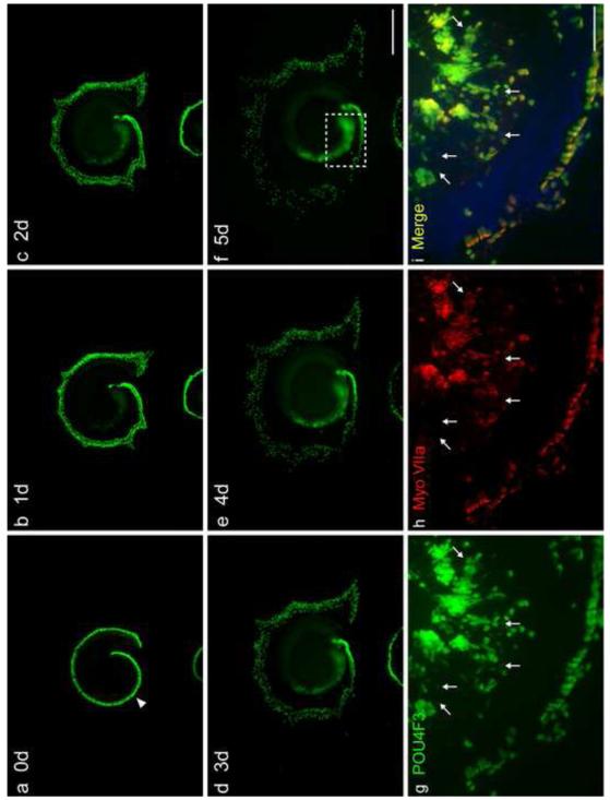
Fig. 7. TF co-transfection enhances Myo7A induction in the GER by ATOH1.
Time course of Pou4f3/GFP expression in the GER of a middle and basal turn explant transfected with hATOH1 plus hTCF3 plus hGATA3, and myosin VIIa expression in the same sample. GER cells showed increasing expression of Pou4f3/GFP from Day 1 to 5, while Pou4f3/GFP+ HCs spread outward and many outer HCs were lost (a-f). Panels g-i show a higher magnification of Pou4f3/GFP, and myosin VIIa expression, for the region indicated in panel f on Day 5. Many Pou4f3/GFP+ GER cells (g) were also positive for myosin VIIa (h, i). While no myosin VIIa+ cells were negative for Pou4f3/GFP, not all Pou4f3/GFP+ GER cells expressed myosin VIIa. Pou4f3/GFP+ cells negative for myosin VIIa tended to show weaker GFP intensity (arrows in g-i indicate some such cells). The scale bar in f = 500 μm. The scale bar in i = 100 μm.

