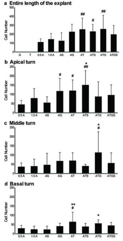
Fig. 9. Quantitative analysis of Myo7A induction by TF co-transfection.
Comparison of the number of Pou4f3/GFP+ GER cells that also expressed myosin VIIa+, observed after transfection with hATOH1 alone or TF combinations. The numbers of Pou4f3/GFP+/myosin VIIa+ GER cells in the entire length of the sensory epithelium (a), in the apical turns (b), in the middle turn (c), and in the basal turn (d) are shown separately. While hATOH1 alone induced Pou4f3/GFP+/myosin VIIa+ GER cells, hTCF3 alone or hGATA3 alone did not. However, co-transfection of each of these TFs with hATOH1 induced more Pou4f3/GFP+/myosin VIIa+ GER cells than hATOH1 alone in the apical turn (a). In the basal turn only hTCF3 enhanced the efeects of hATOH1, while in the middle turn both hTCF3 and hGATA3 were required. The result suggests a synergistic effect among hATOH1, hTCF3, and hGATA3 in the regulation of myosin VIIa expression.
Indicators and abbreviations are the same as those in Fig. 8.

