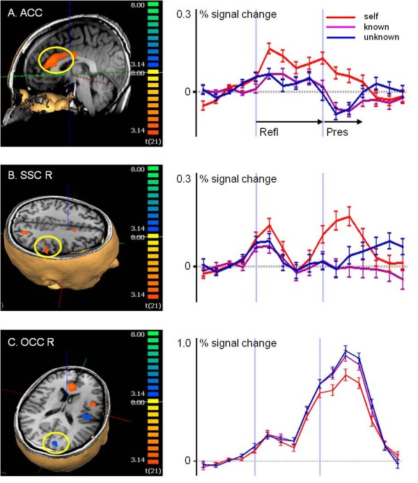
Figure 3.

Brain activity during perception of pictures of oneself. Analysis of selected regions according to the contrast self-perception with greater activity than under known and unknown perception (p < 0.005, Monte Carlo Simulation with cluster wise correction for multiple comparisons p < 0.01). (A) ACC and (B) IPL/SSC R, which were also active during self-reflection. (C) The occipital visual cortex region was less active during self-perception than under the other conditions.
