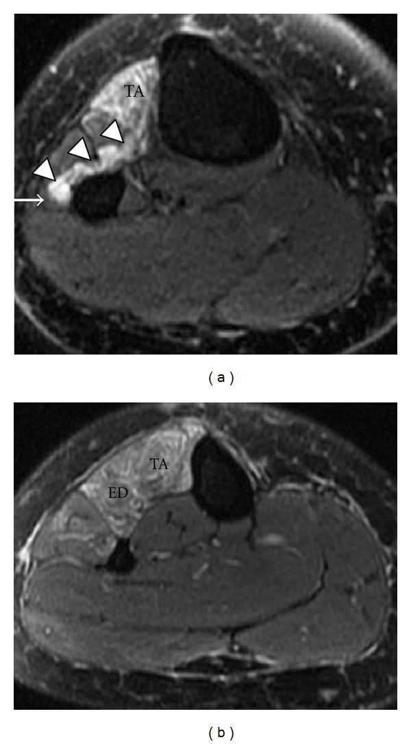
Figure 18.

Common peroneal nerve entrapment secondary to a surgically proven intraneural ganglion cyst in a 44-year-old patient with a 6-month history of right foot drop. Axial T2-weighted fat-saturated images (a, b) reveal a multilobulated high T2 signal structure (arrowheads) compressing the adjacent common peroneal nerve (arrow). Associated patchy high signal in tibialis anterior (TA) and extensor digitorum longus (ED) muscles.
