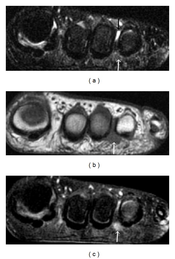
Figure 22.

Morton neuroma in a 38-year-old patient. Coronal T2-weighted with fat-saturation (a), T1-weighted (b), and T1-weighted fat-saturated postcontrast (c) images identify an enhancing tear-drop-shaped soft tissue mass (white arrows) with intermediate signal on both T1- and T2-weighted images in the third intermetatarsal space. A small amount of fluid (black arrow) is noted within the intermetatarsal bursa dorsal to the neuroma in (a).
