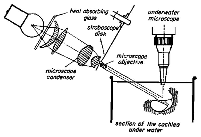
Figure 2.

A section of the cochlea is shown in vitro under water, while illuminated by a stroboscopic lighting system on the left. The observation microscope is vertical and has a long working distance water immersion lens. Note the plane of the baslilar membrane has a tilted and small angle with respect to horizontal. Apparently this angle allowed silver grains scattered onto the BM to be seen as lines when the stroboscopic illumination smeared out their motion (von Békésy, 1960).
