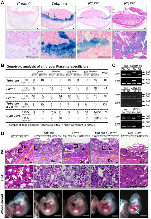
Figure 3. Extra-embryonic Functions of E2F7 and E2F8 are Essential for Embryonic Survival.
(A) X-gal staining showing lineage specific expression of cre recombinase in Tpbp-cre, Plfcre/+ and Pl1cre/+ mice heterozygous for Rosa26loxp reporter allele. (B) Genotypic analysis of embryos derived from intercrosses of Tpbp-cre, Plfcre, Pl1cre and Cyp19-cre with E2f7loxp/loxp;E2f8loxp/loxp mice. For the Tpbp-cre, Pl1cre & Plfcre experiment, cre refers to presence of both cre alleles in heterozygosity. (C) Representative PCR genotyping analyses of E2f7 and E2f8. Genomic DNA was isolated from E10.5 whole placentas (Pla) or laser capture microdissected spongiotrophoblasts (ST) or giant cells (TGC) in Cyp19-cre;E2f7loxp/loxp;E2f8loxp/loxp (Cyp19-cre), Tpbp-cre;E2f7loxp/loxp;E2f8loxp/loxp (Tpbp-cre) and Plfcre/+;E2f7loxp/loxp;E2f8loxp/loxp (Plfcre/+) respectively, along with cre negative controls (con) and whole fetuses (Fet). (D) Representative low and high magnification images of H&E stained E10.5 placental sections (top two rows) and gross appearance of associated fetuses (bottom) with the indicated genotypes. Arrows indicate dilated blood vessels and hemorrhagic areas. Histology scale bars, 100 μm; whole mount scale bars, 1 mm. De., Decidua; La., Labyrinth. Yellow dotted line demarks junctional zone from decidua.

