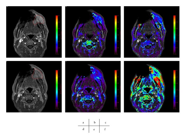
Figure 5.

A poor tumor response to CRT, with an Ohboshi and Shimosato classification of I. The analyses were performed using a proprietary software program (PRIDE software, Philips Healthcare, Eindhoven, The Netherlands). Pre-CRT (a–c), Gd-enhanced T 1 WI (a), K trans map (b), v e map (c), Post-CRT (d–f), Gd-enhanced T 1 WI (d), K trans map (e), v e map (f). The tumor ROIs are delineated in red (a, d). The pre-CRT K trans was 0.13 and K trans decreased after CRT with an average of 0.10. v e decrease from 0.28 to 0.23.
