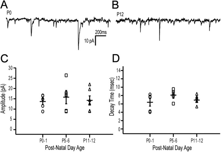
Figure 2.
VSNs form functional synapses with mitral cells before birth. A, Example recording of mEPSCs from a P0 mitral cell. B, Example recording from a P12 mitral cell. All cells were held at −90 mV to amplify events, and events were recorded in the presence of 10 μm gabazine. C, Distribution of the average amplitudes of events at three different age groups (P0–1, n = 5, 13.7 ± 1.52 pA; P5–6, n = 5, 15.8 ± 3.23 pA; P11–12, n = 6, 14.2 ± 2.20 pA). Each value was the average amplitude for a given cell from a minimum of one and a half minutes of data. D, The average decay time for all the events of each cell at each of the three different age groups (P0–1, n = 5, 6.4 ± 0.92 ms; P5–6, n = 4, 8.1 ± 0.75 ms; P11–12, n = 6, 6.9 ± 0.43 ms).

