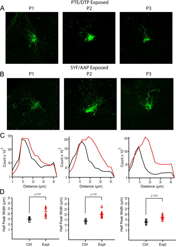
Figure 6.

Activity significantly affects axonal exuberation and coalescence in the AOB. A, Example confocal image stacks of glomeruli across three ages in water exposed control animals. B, Example confocal image stacks of glomeruli from peptide-exposed animals. C, Histogram distributions using Sholl analysis for the two example glomeruli above each histogram (Black, control animal; red, exposed animal). D, Quantification of the half peak width from each histogram across animals at three different ages comparing water controls (Black squares; P1, n = 5, 15.2 ± 1.2 μm; P2, n = 9, 13.9 ± 1.4 μm; P3, n = 9, 12.8 ± 0.7 μm) with exposed animals (Red triangles; P1, n = 6, 23.6 ± 1.7 μm, p = 0.0029; P2, n = 8, 21.7 ± 2.1 μm, p = 0.0076; P3, n = 9, 23.6 ± 1.1 μm, p = 0.001). Scale bar, 20 μm.
