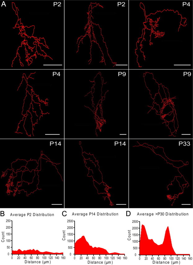
Figure 7.

Mitral cell dendritic morphology undergoes dramatic refinement during the first four postnatal weeks. A, Neurolucida reconstructions of mitral cells across development (P2–33). Mitral cells were targeted, patched, and filled with neurobiotin. Slices were fixed post hoc and stained to be traced and reconstructed using Neurolucida. B–D, Point ending distributions across development. Neurolucida tracings were used and each point ending within the glomerular layer was identified. The histogram of all the distances between each point ending was calculated and averaged for mitral cells from different age groups. P2–4 (B, n = 4), P9–14 (C, n = 4), and P30 and older (D, n = 4). Scale bars, 50 μm.
