Figure 5.
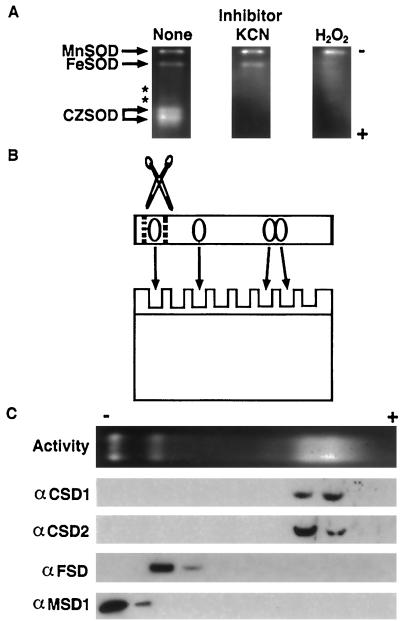
Characterization of the major Arabidopsis SOD activities. A, Forty micrograms of total protein from Arabidopsis rosette tissue was fractionated on a nondenaturing PAGE gel and stained for SOD activity (clear gel regions). Gels were preincubated with KCN (which inhibits CuZnSOD) or H2O2 (which inhibits both CuZnSOD and FeSOD) to facilitate identification of the different activities. Asterisks mark the location of two minor activities that were seen only occasionally. B, Graphic representation of the experiment shown in Figure 5C. SOD activity gels were cut into slices, the slices were boiled in SDS loading buffer to elute and denature proteins, and aliquots were run on SDS-PAGE and immunodetected. C, Immunoblots of SOD activity gel fractions as illustrated in B. Antisera used are listed to the left.

