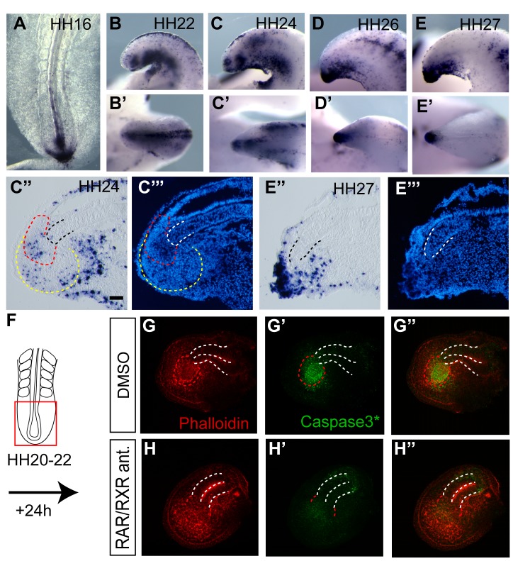
Figure 8. Regulation of cell death in the tailbud by retinoid signalling.
(A–E′, C″, E″) Detection of cell death using Tunel (Apoptag) in chick tailbud at key stages (A) HH16, (B, B′) HH22, (C–C′″) HH24, and (D–E′″) HH26/27. (C″) Some apoptotic cells are detected in mesoderm progenitors (yellow dashed line) and the CNH (red dashed line), distal notochord (black dashed line) at HH24. (C′″) section as in (C″) DAPI stained nuclei confirm tissue organisation. (E″) Increase in apoptosis in the terminal structures by HH27, distal notochord (black dashed line). (E′″) section as in E″ DAPI stained nuclei. (F–H″) Cell death detection using NucView 488 caspase-3 substrate (green) and actin cytoskeleton counter-labelling with Phalloidin (red) in HH20 explanted tailbuds cultured for 24 h in (G–G′) control DMSO only conditions or in (H–H″) the presence of RAR/RXR antagonists. White dashed line, neural tube outline; red dashed line, CNH. CNH region is not well defined in RAR/RXR treated tails. This may reflect ectopic/increased Bra in these conditions. Scale bar, 100 µm.

