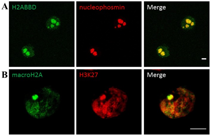
Figure 2. Subnuclear localisation of macroH2A and H2A.Bbd in HeLa S3cells.
HeLa S3 cells stably transfected with either the pOZ -macroH2A1.1 (a) or pOZ- H2A.Bbd (b) plasmid and stained with antibodies against FLAG (green) and H3k27Me3 or nucleophosmin (red). a: macroH2A1.1 is preferentially localised to a single region in interphase HeLa cells and coinsides with H3k27Me3 staining (red). b: H2A.Bbd has a punctate nuclear staining with an exclusion zone (marked by a white arrow) and a strong perinucleolar staining (nucleophosmin, red). The figure shows representative confocal sections. Scale bar: 5 µm.

