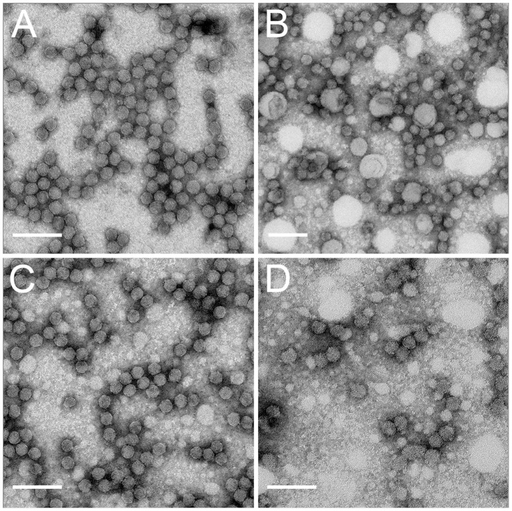
Figure 1. Negative stained grids, coated with CYDV-RPV coat protein antibody, of purified virus from each virus preparation after purification and virus recovered from membranes fed on by R. padi for a 24 h AAP.
Virion morphology was similar within each group and a representative picture for each is shown. Transmissible virions after purification (A) and after membrane feeding (B) look morphologically similar. Non-transmissible virions after purification (C) and after membrane feeding (D) look morphologically similar to each other and are indistinguishable from transmissible virions in shape and size. Scale bars = 100 nm.

