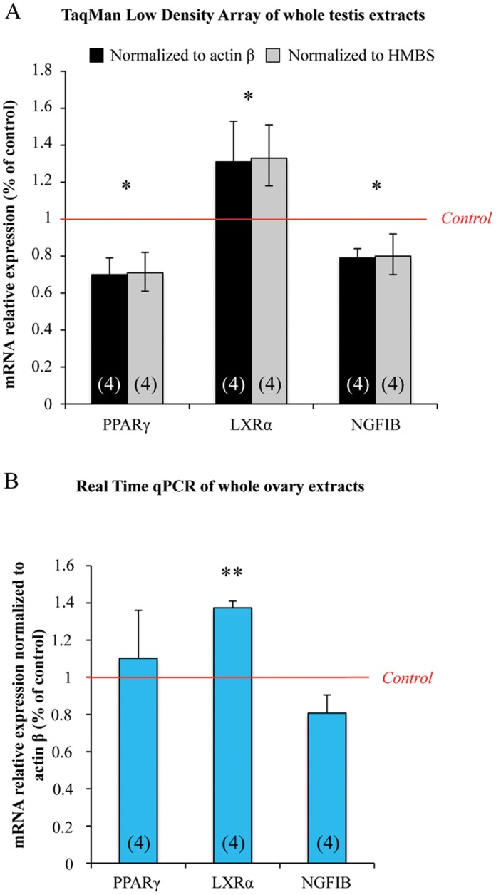
Figure 1. MEHP exposure affects the expression of nuclear receptors in human fetal testes and ovaries.
Testes and ovaries from 7 to 12 week-old human fetuses were cultured with or without 10−4 M MEHP for 3 days and then mRNAs were isolated from whole gonad. (A), NRs superfamily TLDA plates were run with whole testis samples. Results were normalized to actin β or HMBS expression and the 3 differentially expressed NRs are shown as fold changes relative to the control values. Histograms represent the mean ± standard error of the results using 4 independent fetal testis cultures. (B), Transcriptional level of PPARγ, LXRα and NR4A1 was analyzed by real-time qPCR in fetal ovaries. Results were normalized to Actin β expression and are shown as fold changes relative to control values. Histograms represent the mean ± SEM of 4 different ovaries from different fetuses (as indicated in the respective column). *p<0.05, **p<0.01 in paired t-tests recommended when comparing few samples.

